A total of 80 biopsy (skin and blubber) samples were collected. Biopsy samples were collected from 78 individual Bryde’s whales, including six mothers and calves: samples were collected from both the mother and calf in four encounters, the mother only in one encounter, and the calf only in one encounter. A solitary blue whale was sampled, as well as one killer whale (the male in the pod of three individuals).
In 2014, three photographers photographed each close approach: Mizroch photographed from the bow deck and shot at 10 frames per second to try to capture the exact “hit” position of each biopsy dart and to opportunistically capture any photo-ID shots; Jess Taylor photographed from a platform above the upper bridge to capture photo-ID shots and document biopsy hit positions; and Koji Matsuoka photographed from the crow’s nest to capture full body shots of whales, as well as biopsy hit locations and photo-ID shots.
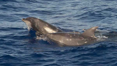
Figure 2. Bottlenose dolphins in the pelagic North Pacific. Photo by Sally Mizroch.
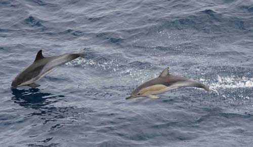
Figure 3. Common dolphins: normal pigmentation (right) and a color morph described by Perrin in 1995 (left). Photo by Sally Mizroch.
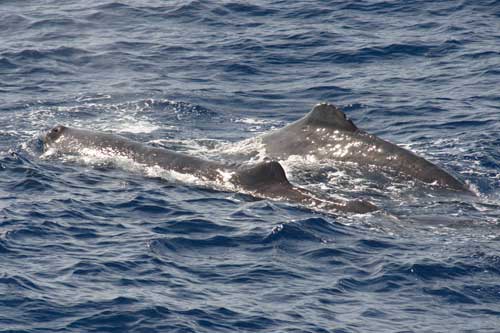
Figure 4. Sperm whale mother and calf. Photo by Sally Mizroch.
In 2014, as in 2011 and 2012 (surveys in which Mizroch also participated), all of the POWER photos were geotagged; however, in 2014, the photos were geotagged automatically using a GPS logger attached directly to each camera.
At the end of each survey day, photos from all three cameras were integrated and transferred to folders labelled with the day’s date and line-transect sighting number. An informative encounter number that included information abouth the cruise (POWER) and the ship (YS3) plus the sighting number, for example, 2014_POWER_ YS3_20140731_23, was batch edited into the “Image Description” EXIF metadata field of each photo in each folder.
Photographs were evaluated each evening to determine if there was sufficient detail to assign a catalog number to individual whales. All cataloged photographs were labelled with an annual catalog number in the “Artist” EXIF metadata field, and the body part used to identify the individual (e.g., LD for left dorsal) was typed into the “Copyright” EXIF metadata field. For each successful biopsy encounter, photos of the biopsy hit were labelled with the biopsy sample number and shooter name, and then the photos were evaluated jointly with the biopsy sample manager to determine the “hit position” to enter on the biopsy data form.
When all the photo labelling was complete, the daily photographs were backed up each night to three hard drives using a high-speed, hard-drive dock (155 Mb/sec). Each photographer was able to review all of the integrated photos each day and then adjust camera settings (ISO, shutter speed, zoom level) in subsequent photos, if necessary, to complement photos taken by the other photographers.
In addition to the many thousands of spectacular Bryde’s whale photos taken during the survey (Fig. 1), a number of other interesting species were documented in the middle of the North Pacific: for example, three groups of bottlenose dolphins in a pelagic region far from other known sightings of this species (Fig. 2); a number of common dolphin groups, including some of the classic color morphs described by Perrin in 1995 (Fig. 3); and a sperm whale mother and calf (Fig. 4).